AS Level Biology 9700
1. Cell Structure
Written by: Pranav I
Formatted by: Pranav I
Index
1.1 Cells are the basic units of life
1.2 Cell biology and microscopy
1.3 Plant and animal cells as seen with a light microscope
1.4 Measuring and calculating magnification
1.5 Electron microscopy
1.6 Plant and animal cells as seen with an electron microscope
1.7 Bacteria
1.8 Comparing prokaryotic cells with eukaryotic cells
1.9 Viruses
1.1 Cells are the basic units of life
- Cells are surrounded by a thin partially permeable membrane which controls exchange between the cell and its environment
- Eukaryotic cells contain membrane-bound nuclei with the genetic material, DNA (e.g. animals, plants, fungi)
- Prokaryotic cells lack membrane-bound nuclei and the DNA is free in the cytoplasm (e.g. bacteria)
1.2 Cell biology and microscopy
- Cells are examined and studied using microscopes
- Light microscopes → use light as the source of radiation
- Electron microscopes → use electrons as the source of radiation
- Micrographs: a picture taken with the aid of a microscope
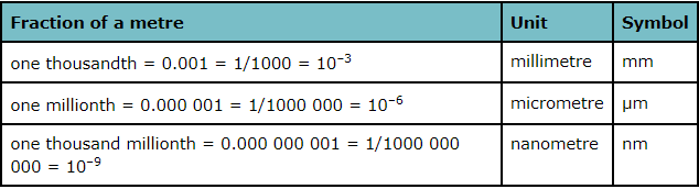
- Cells vary in size from about 5 µm to 40 µm
- Average bacterial cell → 1 µm across
- Diameter of ribosomes → 25 nm
1.3 Plant and animal cells as seen with a light microscope
- Both the eyepiece lens and the objective lens magnify the image
- A photomicrograph is a photograph of a specimen as seen with a light microscope
- Most cell contents are colorless and need to be stained with colored dyes
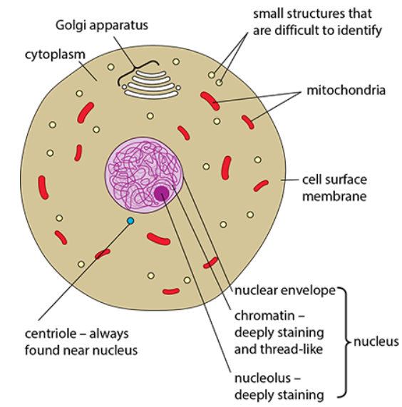
Features that animal cells and plant cells have in common
- Cell surface membrane
- Referred to as the plasma membrane
- Partially permeable → controls the exchange of materials between the cell and its environment
- Found in ALL cells
- Nucleus
- Chromatins → the material of which chromosomes are made containing DNA, proteins, and small amounts of RNA (a mass of coiled threads)
- Chromosomes are formed from tightly coiled chromatin
Nucleolus → made of loops of DNA; deeply staining; the main function is to make ribosomes, and one to five are found in mammals
- Cytoplasm
- Protoplasm (all the living material inside the cell) = Cytoplasm + Nucleus
Aqueous material (fluid to jelly-like consistency) in which many organelles can be found - Organelles are functionally and structurally distinct parts of the cell that are often surrounded by one or two membranes
- Cell activities are organized into compartments for efficiency
- Protoplasm (all the living material inside the cell) = Cytoplasm + Nucleus
- Mitochondria
- Specialised to carry out aerobic respiration
- Most numerous organelles seen with the light microscope
- Golgi apparatus
- Collects and processes molecules within the cell, particularly proteins
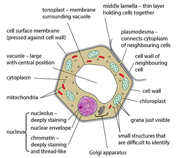
Differences between animal cells and plant cells
- Centrioles
- Small structures close to the nucleus
- Found only in animal cells
- Cell walls and plasmodesmata
- Plant cells are surrounded by a cell wall
- Relatively rigid due to fibers of cellulose
- Gives the cell a definite shape
- Prevents the cell from bursting when water enters by osmosis
- Freely permeable → allows free movement of molecules and ions
- Plasmodesmata are pores containing fine strands of cytoplasm → link neighboring cells
- Lined with the cell surface membrane
- Vacuoles
- Plant cells have a large, permanent, central vacuole surrounded by the tonoplast
- Regulate osmotic properties of the cell
- Contain pigments
- Animal cells have small, temporary vacuoles only
- Chloroplasts
- Specialized for photosynthesis
- Grana: stacks of membranes containing chlorophyll inside a chloroplast
1.4 Measuring and calculating magnification
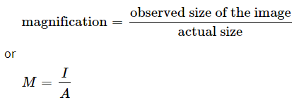
- Magnification is the number of times larger an image of an object is than the real size of the object
Measuring cell size
- Eyepiece graticule: a small, transparent scale that can be inscribed in the microscope eyepiece (usually has 100 divisions)
- However the eyepiece graticule needs to be calibrated
- Stage micrometer: Very small, accurately drawn scale of known dimensions, engraved on a microscopic slide → commonly has subdivisions of 0.1 and 0.01 mm
- Used to calibrate the eyepiece graticule
- Superimposing both of the scales will help determine the cell’s size
1.5 Electron microscopy
Magnification and resolution
- Resolution: the ability to distinguish between two objects very close together; the higher the resolution of an image, the greater the detail that can be seen
- Maximum resolution of a light microscope: 200 nm
- Note: resolution does not increase with magnification
The electromagnetic spectrum
- Wavelengths of visible light: 400 nm (violet) to 700 nm (red)
- The limit of resolution is half the wavelength of the radiation used
- Transparent objects have to be stained to be seen
The electron microscope
- Free electrons behave like EM radiation → have a very short wavelength
- Suitable for microscopy due to their short wavelength and negative charge (can be easily focused using electromagnets)
- Maximum resolution of a transmission electron microscope: 0.5 nm
- Maximum resolution of a scanning electron microscope: 3 nm to 20 nm
Transmission and scanning electron microscopes
- Transmission electron microscopes (TEM)
- The beam of electrons is passed through a thin section of the specimen
- Only the electrons that are transmitted (pass through) are seen
- Can see inside cells
- Scanning electron microscopes (SEM)
- The beam of electrons is used to scan the surfaces of structures
- Only the reflected beam is observed
- Surface structures can be seen
Viewing specimens with the electron microscope
- The electron beam is projected onto a fluorescent screen (electrons cannot be seen)
- Gives a black and white picture
- Stains containing heavy metal atoms are used to improve contrast (stop the passage of electrons)
- False-colour images are created using a computer
- The electron beam, specimen and the fluorescent screen must be in a vacuum
- All specimens must be dehydrated since water boils at rtp in vacuum
1.6 Plant and animal cells as seen with an electron microscope
- Ultrastructure: the fine, detailed structure of a cell as revealed by the electron microscope
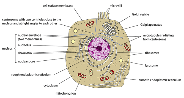
- Cell surface membrane
- Extremely thin (7 nm)
- Partially permeable and controls exchange between the cell and its environment
- Contains three layers → two dark layers with a pale interior
- Microvilli
- Small, finger-like extensions of the CSM
- Increase the surface area for more efficient absorption or secretion
- Nucleus
- Largest cell organelle
- Nuclear envelope
- The two membranes, situated close together, that surround the nucleus
- The outer membrane is continuous with the ER
- Nuclear pores
- Pores found in the nuclear envelope which control the exchange of materials between the nucleus and the cytoplasm
- Nucleus contains chromosomes
- DNA molecules combine with histone proteins → chromatin (fold up into a compact shape)
- Chromatin also has some RNA
- The nucleolus makes ribosomes → contains genes that code for ribosomal RNA (rRNA)
- rRNA is combined with ribosomal proteins to make ribosomes
- Endoplasmic reticulum
- A network of flattened sacs (cisternae) running through the cytoplasm of eukaryotic cells
- Molecules can be transported through the cell inside the sacs separate from the cytoplasm (especially proteins)
- The rough ER is covered with ribosomes
- The smooth ER makes lipids and steroids; it is also a major storage site for calcium ions
- Ribosomes
- Consist of a large and a small subunit
- Eukaryotes have 80S ribosomes
- Svedberg (S) units: Measure of how rapidly substances sediment in an ultracentrifuge → higher the value, faster the sedimentation
- Made of ribosomal RNA and protein
- Allow all molecules involved in protein synthesis to gather in one place
- Golgi apparatus
- A stack of flattened sacs called cisternae, constantly forming at one end and breaking up into Golgi vesicles at the other end
- Collects and processes molecules, particularly proteins from the RER
- Processed molecules are transported to other parts of the cell in Golgi vesicles
- Golgi vesicles are used to make lysosomes
- Sugars are added to proteins to make glycoproteins
- Sugars are added to lipids to make glycolipids
- Golgi enzymes synthesize new cell walls during plant cell division
- Release mucin in goblet cells
- Lysosomes
- 0.1-0.5 µm in diameter
- Surrounded by a single membrane
- Contain hydrolases to break unwanted substances and structures (old organelles or entire cells)
- Contents of lysosomes are acidic (suitable for hydrolysis)
- Engulf and destroy unwanted cell components
- Fuse with endocytic vacuoles and digest their contents
- Lysosomal enzymes are released from the cell for extracellular digestion
- Autolysis (digestion of the cell)
- Mitochondria
- 1 µm in diameter
- Surrounded by two membranes (mitochondrial envelope)
- Space between two membranes: intermembrane space
- Cristae: folds of the inner membrane of the mitochondrial envelope
- Matrix: interior of the mitochondrion
- Carry out aerobic respiration to produce ATP molecules (universal energy carrier)
- ATP is broken down into ADP to release energy
- Have 70S ribosomes and circular DNA molecules
- Endosymbiont theory → mitochondria and chloroplasts are ancient bacteria living inside larger eukaryotic cells
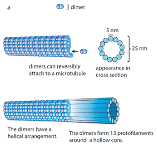
- Microtubules and microtubule organizing centres (MTOCs)
- 25 nm in diameter
- Make up the cytoskeleton which determines cell shape
- Made of a protein called tubulin
- Two forms → α-tubulin and ß-tubulin
- α- and ß-tubulin molecules combine to form dimers
- Dimers join end to end to form long protofilaments
- Thirteen protofilaments lined up alongside each other in a ring to form a microtubule
- Secretory vesicles and other cell components can be moved along the surface of the MTs → intracellular transport system
- A spindle made of MTs is used to separate chromatids during nuclear division
- Form part of the structure of centrioles
- Essential part of the mechanism involved in the beating movements of cilia and flagella
- MTOCs control the assembly of MTs
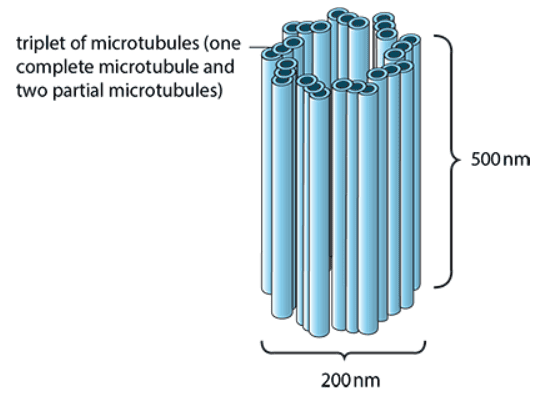
- Centrioles and centrosomes
- 200 nm in diameter; 500 nm long
- Two centrioles present just outside the nucleus of animal cells
- Lie in a region known as the centrosome
- Absent from most plant cells
- Each contains nine triplets of microtubules
- Centrosomes assemble MTs to make up the spindle during nuclear division
- Centrioles are needed for the production of cilia
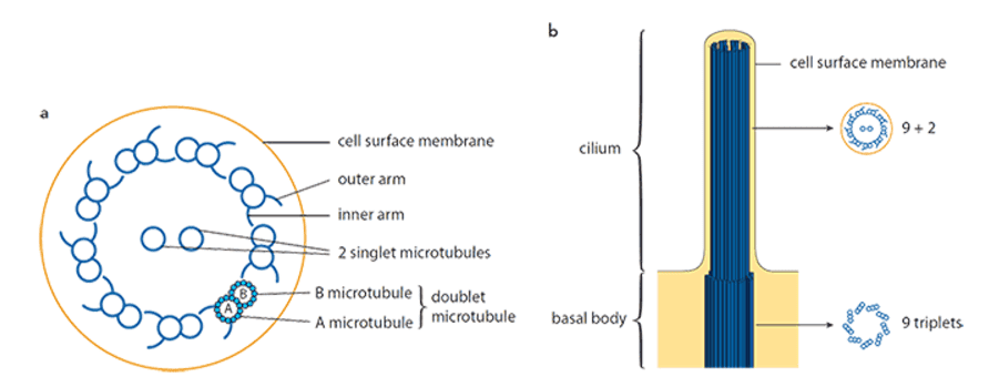
- Cilia and flagella
- Whip-like, beating extensions
- Surrounded by an extension of the CSM
- Cilia → short and numerous ; Flagella → long and 1-2 per cell
- Cilia have 2 central MTs and a ring of nine MT doublets (MTDs) around the outside → ‘9 + 2’ structure
- Each MTD has an A microtubule and a B microtubule → A is a complete ring of 13 protofilaments; B is an incomplete ring of 10 protofilaments
- Each A MT has inner and outer arms made of dynein → make contact with and move along neighboring B MTs during beating
- Sliding motion converted to bending by other parts
- Basal body (identical to centrioles) present at the base of each cilium
- Centrioles replicate to produce basal bodies, from which cilia and flagella grow
- Maintain a flow of fluid (e.g: Mucus in the respiratory tract)
- Useful for the locomotion of single-celled organisms
- Chloroplasts
- 3-10 µm in diameter
- Surrounded by the chloroplast envelope
- Chlorophyll is found on the membranes of chloroplasts
- Carry out photosynthesis
- Thylakoids (stack to form grana)
- Flattened, membrane-bound, fluid-filled sacs which are the site of the light-dependent reactions of photosynthesis
- Light-independent reactions occur in the stroma
- Sugars produced are stored as starch grains
- Lipid droplets for making membranes found in the stroma
- Have 70S ribosomes and circular DNA
- Endosymbiont theory applies
- Cell walls
- Primary wall is formed first
- Relatively rigid → cellulose fibres are inelastic and have high tensile strength
- Parallel fibres of cellulose running through other polysaccharides
- Secondary wall is formed next
- Cellulose fibres of different layers run in different directions → stronger cross-ply structure
- Lignin is added for additional strengthening in some cell walls → provides compressional strength
- Mechanical strength and support for cells and the plant → lignification and turgid tissues
- Cell walls prevent cells from bursting by osmosis
- Interconnected cell walls form the apoplast pathway for transport
- Plasmodesmata form the symplast pathway for transport
- Suberin in root endodermal cell walls to block transport in the apoplast pathway
- Cutin in epidermal cell walls to reduce water loss by evaporation
- Vacuoles
- Solution in the vacuole is relatively concentrated
- Water enters vacuoles by osmosis and causes a build-up of pressure → turgid tissues help support non-woody stems
- May contain hydrolases and act as lysosomes (in plants)
- Store secondary metabolites
- Act as food reserves
- Store waste products
- Growth in size of plant cells due to the osmotic uptake of water into the vacuoles
1.7 Bacteria
- Prokaryotes with simpler cells → 1000 times smaller and lack a nucleus surrounded by a double membrane
Structure of bacteria
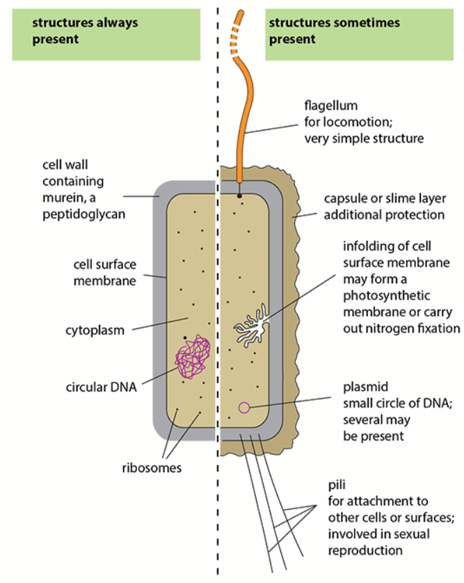
- 1-5 µm in diameter
- Cell wall
- Contains murein, a peptidoglycan, for more rigidity
- Prevents the cell from bursting when water enters the cell by osmosis
- Cell surface membrane
- Cytoplasm
- Does not contain any double membrane-bound organelles
- Circular DNA
- Found in a region called the nucleoid along with proteins and small amounts of RNA
- The DNA molecule is circular (closed loop with no free ends)
- 70S ribosomes
- Flagellum
- Simple hollow cylinder made of identical protein molecules
- Rigid, wave-shaped structure → rotates at its base to propel the bacterium
- Infolding of the CSM
- For certain biochemical reactions to take place
- In blue-green bacteria → photosynthesis (contains photosynthetic pigments)
- In nitrogen fixing bacteria → nitrogen fixation (converting atmospheric nitrogen to nitrogen-containing compounds)
- Capsule or slime layer
- Extra layer outside the cell wall → protect from drying out
- Capsule → definite structure made of polysaccharides
- Slime layer → more diffuse and easily washed off
- Plasmid
- Small circle of DNA separate from the main DNA
- Contains few genes → give resistance to particular antibiotics
- Can copy themselves independently
- Numerous in a cell
- Pili
- Fine protein rods → 1-100 on the outside of a cell
- Attachment and interactions with other cells and surfaces
- Transfer of genes from one bacterium to another during conjugation
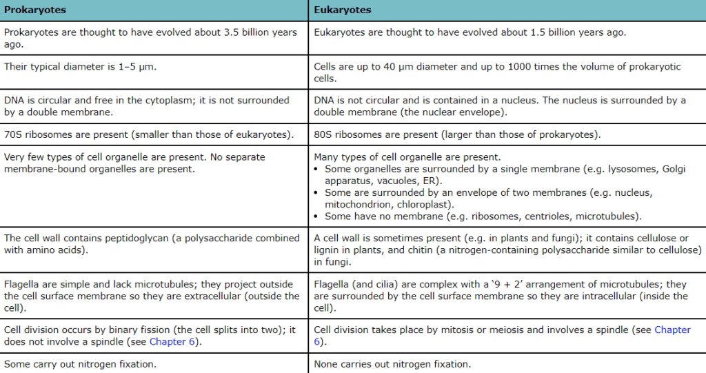
1.8 Comparing prokaryotic cells with eukaryotic cells
1.9 Viruses
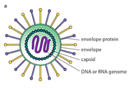
- 20-300 nm in diameter
- Do not have a cellular structure
-
Consist of:
- A self-replicating molecule of DNA or RNA
- A protective coat of protein molecules (capsomeres) called a capsid
- A membrane-like envelope made of phospholipids (proteins may project from it)
- Parasitic since they can only reproduce by infecting and taking over living cells